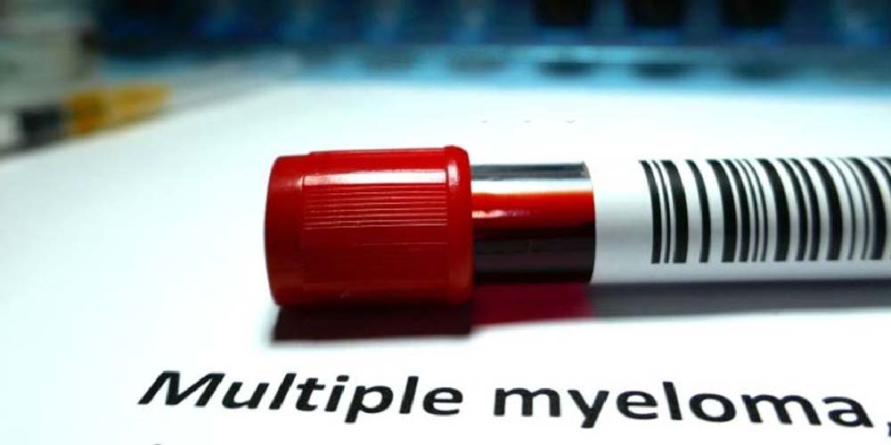
DIAGNOSIS & EVALUATION OF MULTIPLE MYELOMA
Dr. Gaurav Dixit is an esteemed International Hemato- oncologist (Blood Cancer Specialist). He has been awarded as a “Professional Certification in Multiple Myeloma” by Mayo Clinic- USA.
Dr Gaurav Dixit is well versed with advances in the field of Haematology and always follows the latest international guidelines for deciding better treat plan for his patients by keeping himself updated.
Due to an exceptional understanding about Hemato-Oncology, Dr Gaurav Dixit is always willing to interact, learn and help. This interactive blog enlightening the diagnostic workup road map for evaluation of Multiple Myeloma by him is need of the hour.
Myeloma is the blood cancer of plasma cells present in the bone marrow.
How Myeloma does begin? – The Myeloma begins as healthy plasma cells start changing and grow out of control. This also results in multiple bone lesions that’s where the phrase "multiple myeloma" exists.
The abnormal plasma cells start crowding out & also suppress the growth of other cells in the bone marrow. With the reduced creation of normal plasma cells, a person’s immunity is compromised.
The compromised immunity state in patients of multiple myeloma is associated with:-
- Anemia due to shortage of red blood cells
- Excessive bleeding from cuts to the skin as there is a shortage of platelets
- Decreased ability to fight infection due to deprivation of white blood cells
- The host’s inability is not uncommon to respond to infection because of the presence of abnormal antibodies
It is pertanious here to mention that like regular plasma cells, myeloma cells can produce antibodies. But myeloma cells are unable to produce healthy &/or functioning antibodies.
Myeloma cells make “monoclonal protein,” "monoclonal immunoglobulin," or “M protein.”
M protein builds up in the blood and urine, potentially damaging the kidneys and other organs, & is also responsible for reducing immunity.
Myeloma results in structural bone damage, leading to weakened bones and painful fractures.
On hematological examination microscopically, the multiple myeloma is characterized by numerous malignant plasma cells. These cells are characterized by a pale area within the cytoplasm, near the nucleus comparable with normal bone marrow cells.
Dr Gaurav Dixit (well-knowned Hemato-oncologist) performs well planned cost effective interdisciplinary diagnostic workup to evaluate/diagnose, multiple myeloma. Besides this Hemato-oncologist also test to learn in case cancer has spread to another part of the body from where it started and this is referred to as staging; this allows deciding fast and save treatment plan. This also allows him to ensure better survival rate for his patients.
Well! For almost all cancer a biopsy is the only golden diagnostic pavement to confirm cancer along with its typing or subtyping.
During a procedure called a biopsy, small sample of tissue for testing in a laboratory or pathology lab is taken either by surgery or aspiration cytology. In cases where biopsy is not possible, then by other tests diagnostic workup is accomplished.
Here options for diagnoses and evaluation of multiple myeloma are discussed by Dr Gaurav Dixit. Remember all tests listed below will not be used for every case.
By this evaluation, the hematoncologist will be able to consider the following parameters’ before choosing a diagnostic test:
- The type of cancer suspected
- Your signs and symptoms
- Your age and general health
- The results of earlier medical tests
The enlisted tests can be used to diagnose multiple myeloma:
1. Blood and urine tests:
Myeloma cells secrete the antibody monoclonal immunoglobulin referred to as M protein. Circulating M protein levels in a host’s blood and urine are used to determine the extent of the disease. This also helps in monitoring the effectiveness of treatment.
In certain cases, the myeloma cells only secrete part of the antibody, namely the light chain.
Serum protein electrophoresis (SPE or SPEP) or urine protein electrophoresis (UPE or UPEP):
It is the amount of M protein in the blood or urine on measurement by serum protein electrophoresis (SPE or SPEP) or urine protein electrophoresis (UPE or UPEP) helps to see the disease is progressing or coming back after treatment or treatment is working.
By measuring the immunoglobulin levels are measured to help check the number of antibody levels in the blood. These antibodies are:
a. immunoglobulin G (IgG)b. immunoglobulin A (IgA)
c. immunoglobulin M (IgM)
When the cancer protein level is up in multiple myeloma- the normal antibody levels are down.
Serum-free light chain assay-(more sensitive test than measuring M protein in the urine)
Measuring the number of free light chains in the blood is done before the blood is filtered by the kidneys by the test serum free light chain assay. The light chain found in the urine is called the Bence Jones protein.
By blood tests levels of serum albumin and serum beta-2 microglobulin (β2-M) are also measured.
Serum albumin - blood protein made by the liver necessary for maintaining proper blood volume and general health.
β2-M - a small protein that plays an important role in the body's immune response.
The results of blood tests for levels of serum albumin and serum beta-2 microglobulin (β2-M) are important for determining the stage of myeloma.
2. Imaging/X-ray
Using a small amount of radiation allows imaging - typically the first step in evaluating bones when myeloma is suspected or diagnosed.
3. Magnetic resonance imaging (MRI)
MRI uses magnetic fields to produce detailed images of the body. It also shows if the normal bone marrow has been replaced by myeloma cells especially in the skull, spine, and/or pelvis. The detailed images reveal compression fractures of the spine or a tumor pressing on nerve roots which also enables to measurement of the tumor’s size.
4. Computed tomography (CT or CAT) scan
The cross-sectional view revealing abnormalities or tumors in soft tissues is enabled by CAT. A computer provides a 3-dimensional image of the inside of the body and intravenous contrast dye often used for CT scans for other types of cancer is specifically avoided in multiple myeloma. So on imaging prescription form, it is pertanious for the oncologist to mention a provisional diagnosis of multiple myeloma to alarm before dye injection into a patient’s vein because this can cause kidney damage in people with myeloma.
5. Positron emission tomography (PET) or PET-CT scan
A PET scan is usually combined with a CT scan as a way to create pictures of organs and tissues inside the body. By a small amount of a radioactive sugar, substance injected into the patient’s body which is taken up by cells that use the most energy and because cancer cells tend to use energy actively; it absorbs more of the radioactive substance. A scanner is enabled to detect substances to produce images of the inside of the body.
6. Bone marrow aspiration and biopsy
To examine the bone marrow; as bone marrow has both a solid and a liquid component:
A bone marrow aspiration removes a sample of the fluid with a needle.
A bone marrow biopsy removes a small amount of solid tissue using a needle.
A common site for a bone marrow aspiration and biopsy is the pelvic bone.
A pathologist analyzes the above-mentioned sample(s).
7. Cytogenetics
The genes in the myeloma are examined by cytogenetics. Cytogenetics is special testing referred to as fluorescent in situ hybridization (FISH). Cytogenetics analyzes a cell's chromosomes and determines the genetic makeup of the myeloma and whether it is standard or high risk.
8. Fat pad aspirate
Certain M proteins which are misfolded get deposited in body tissues causing organs to stop functioning normally. This condition is called amyloidosis9. Molecular testing of the tumor
Already mentioned above, tests on a tumor and/or bone marrow sample to identify specific chromosomes (cytogenetics), genes (FISH or genomic sequencing; see above), proteins, and other factors. Results of these tests also help to determine the treatment regimen.
Cytogenetics is performed on a tissue sample removed during a biopsy to find out how aggressive the cancer is. In myeloma, the genes in plasma cells studied using the FISH test to identify standard and high-risk disease; help guide treatment planning.
Minimal residual disease (MRD): As treatments have become efficacious in treating myeloma and reporting follow up prognosis. The new approaches to measure how well a treatment works have been developed, including MRD. If "MRD positive", further treatment may be offered to suppress the disease further; most efficaciously assessed by Flow cytometry. To know more about good prognosis followed by best treatment for Multiple Myeloma, diagnosis and evaluation is first step. For better treatment plan followed by increased disease free survival rate; an appropriate diagnostic pavement is a necessity which is provided by an esteemed Clinical Hemato-oncologist- Dr Gaurav Dixit has an experience of 19 years. Currently practicing in Action cancer hospital,A-4, Paschim Vihar, New Delhi, Delhi, India 110063.
For consultation please see following contact details: Dr Gaurav Dixit DM (clinical hematology) Consultant clinical Hematologist and BMT physician Action cancer hospital A-4 Paschim Vihar, New Delhi 110063 He can be reached at following Website :https://www.drgauravdixit.com